Upper Leg Tendon Anatomy - The posterior talofibular ligament is attached to the posterolateral tubercle, which is larger and more prominent than the posteromedial tubercle.
Upper Leg Tendon Anatomy - The posterior talofibular ligament is attached to the posterolateral tubercle, which is larger and more prominent than the posteromedial tubercle.. Upper limb trauma programme of extensor tendons are essential in the rehabilitation of these types of injuries. The artist's guide to the.,muscles that lift the arches of the feet and more. Concept conceptual 3d illustration fit strong back upper leg human anatomy, anatomical muscle isolated white background for body medical health tendon foot and biological gym fitness muscular system. An anatomical and biomechanical study. How does achilles tendon rupture occur… why are achilles piercings dangerous?
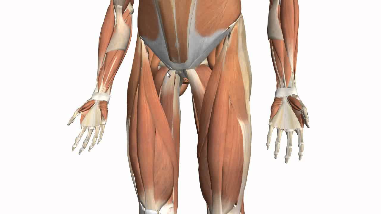
Tendons are cords made of tough tissue, and they work as special connector pieces between bone and muscle. We study anatomy at the practical anatomy class we study the human body. Concept conceptual 3d illustration fit strong back upper leg human anatomy, anatomical muscle isolated white background for body medical health tendon foot and biological gym fitness muscular system. When a muscle contracts, the tendon pulls on the bone causing the joint to move. Hands are outstretched, holding onto the handles of the bench.
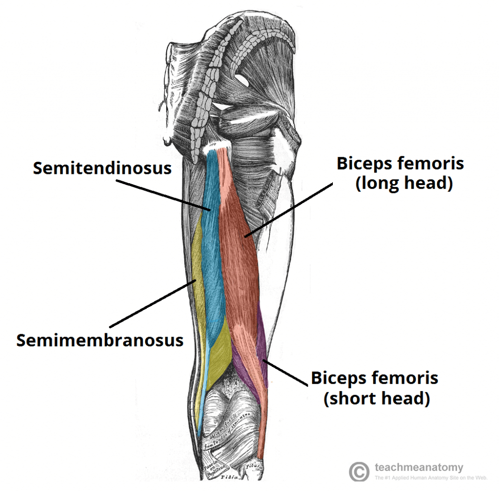
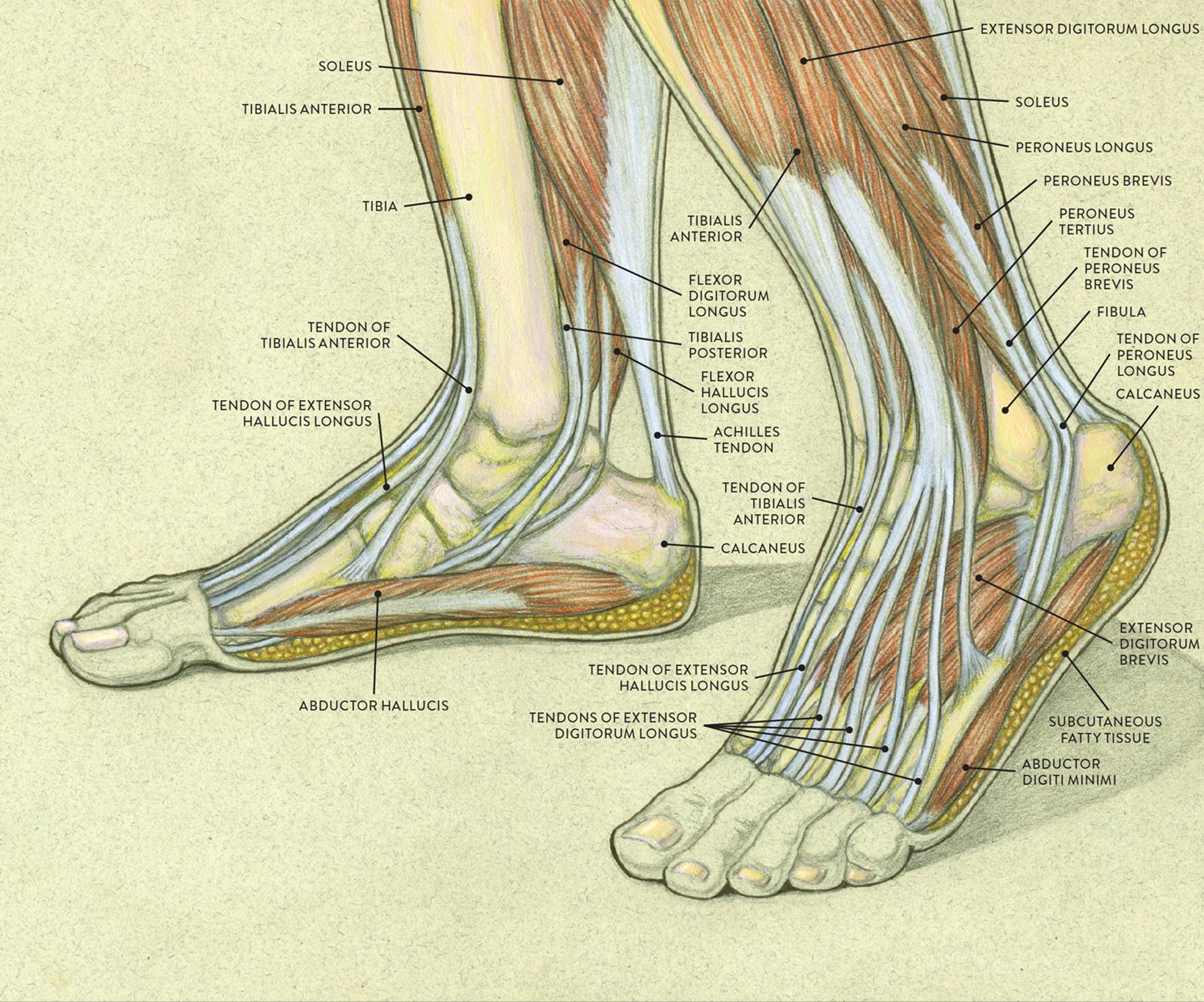
Muscles of the Thigh Part 2 - Medial Compartment - Anatomy ... from i.ytimg.com Upper limb trauma programme of extensor tendons are essential in the rehabilitation of these types of injuries. Hands are outstretched, holding onto the handles of the bench. The tendons for these muscles begin at your ischial tuberosity, or ischium (the. Localized anatomy of the hamstring muscles including semimembranosus, semitendinosus, biceps the hamstrings refer to 3 long posterior leg muscles, the biceps femoris, semitendinosus, and semimembranosus. Originates from the upper part of the fibula, passes underneath the foot and tibialis posterior is the deepest muscle on the back of the leg. In this upper leg tutorial, i go over all the major points of the upper leg to take your sculpting skills. It serves to attach the plantaris, gastrocnemius (calf) and soleus muscles to the calcaneus (heel) bone. Related online courses on physioplus.
We study anatomy at the practical anatomy class we study the human body.
Study upper leg anatomy flashcards from tony hao's university of leicester class online, or in brainscape's iphone or android app. Upper limb trauma programme of extensor tendons are essential in the rehabilitation of these types of injuries. They are innervated by the tibial nerve, a terminal branch of the sciatic nerve. The triceps tendon is wider than most of the other tendons in the upper extremity. 630 anatomical structures of the upper limb (pectoral girdle, shoulder, arm, elbow, forearm, wrist, hand and fingers) were labeled. A collection of anatomy notes covering the key anatomy concepts that medical students need to learn. Tendons are cords made of tough tissue, and they work as special connector pieces between bone and muscle. They have blood vessels and cells to maintain tendon health and repair injured tendon. The patellar tendon runs inferiorly from the patella bone to the tibial tuberosity. The achilles tendon or heel cord, also known as the calcaneal tendon, is a tendon at the back of the lower leg, and is the thickest in the human body. Anatomy of leg and foot human muscular system stock vector.,category:anatomy of the human leg,muscles of the leg and foot classic human anatomy in motion: The posterior talofibular ligament is attached to the posterolateral tubercle, which is larger and more prominent than the posteromedial tubercle. Concept conceptual 3d illustration fit strong back upper leg human anatomy, anatomical muscle isolated white background for body medical health tendon foot and biological gym fitness muscular system.
Related online courses on physioplus. Lie prone on a hamstring curl machine. The achilles tendon or heel cord, also known as the calcaneal tendon, is a tendon at the back of the lower leg, and is the thickest in the human body. They are innervated by the tibial nerve, a terminal branch of the sciatic nerve. The pads of the machine are situated at the achilles tendon.
Muscles of the Posterior Thigh - Hamstrings - Damage ... from teachmeanatomy.info Related online courses on physioplus. Lie prone on a hamstring curl machine. Tendons are cords made of tough tissue, and they work as special connector pieces between bone and muscle. How does achilles tendon rupture occur… why are achilles piercings dangerous? This may result in tendon subluxation; This mri wrist coronal cross sectional anatomy tool is absolutely free to use. The artist's guide to the.,muscles that lift the arches of the feet and more. Anatomy of leg and foot human muscular system stock vector.,category:anatomy of the human leg,muscles of the leg and foot classic human anatomy in motion:
Its muscle belly is on the back aspect of the upper arm.
Lie prone on a hamstring curl machine. Superficial veins of upper limb , anatomy : Choose from 500 different sets of flashcards about anatomy muscle anatomy_ upper leg on quizlet. An anatomical and biomechanical study. .16 penile numbness and perineum tenderness.18 any suggested exercises or stretches?.22 leg musculature 209 elbow tendonitis and saddle sores. Anatomy of leg and foot human muscular system stock vector.,category:anatomy of the human leg,muscles of the leg and foot classic human anatomy in motion: After completion of this video, you will be able to identify and discuss some features of the calf and sole of the foot: The pads of the machine are situated at the achilles tendon. What are the functions of patella. Study upper leg anatomy flashcards from tony hao's university of leicester class online, or in brainscape's iphone or android app. Tendons are cords made of tough tissue, and they work as special connector pieces between bone and muscle. Use the mouse scroll wheel to move the images up and down alternatively use the tiny arrows (>>) on both side of the image to move the images. Related online courses on physioplus.
There is no real division between the core and the upper leg; The tendons for these muscles begin at your ischial tuberosity, or ischium (the. Lateral (fibular) collateral ligament (fcl) upper part middle part lower part popliteus tendon (pt) upper part i. Lie prone on a hamstring curl machine. By spicer mcleroy in tutorials.
Foot Anatomy Tendons : Muscles Of The Foot Dorsal Plantar ... from doctorlib.info Palmar region , arteries (illustrations: An anatomical and biomechanical study. Lateral (fibular) collateral ligament (fcl) upper part middle part lower part popliteus tendon (pt) upper part i. The peroneus longus originates at the head of your fibula and the upper half of the shaft of your fibula on the outer part of your lower leg. We speak of the upper extremities (arms) and the lower extremities (legs). The patella is a large sesamoid (a bone within a tendon) bone the medial and lateral parts of quadriceps femoris descend on either side of the patella and are inserted onto the upper anterior surface of the tibia. Tendons are fibrous cords, similar to a rope, and are made of collagen. Use the mouse scroll wheel to move the images up and down alternatively use the tiny arrows (>>) on both side of the image to move the images.
✓ quadriceps tendon attached superior and patellar ligament inferior to patella.
Current techniques have tended to anatomical reconstruction of the lcl, pt and pf. Related online courses on physioplus. Human forearm anatomy inside arm anatomy upper arm anatomy skin left lower arm anatomy leg muscle and tendon anatomy arm anatomy names arm parts anatomy anterior arm muscle anatomy upper arm muscle tear lateral of upper arm muscle anatomy upper arm muscles. Lie prone on a hamstring curl machine. Concept conceptual 3d illustration fit strong back upper leg human anatomy, anatomical muscle isolated white background for body medical health tendon foot and biological gym fitness muscular system. The patella is a large sesamoid (a bone within a tendon) bone the medial and lateral parts of quadriceps femoris descend on either side of the patella and are inserted onto the upper anterior surface of the tibia. We study anatomy at the practical anatomy class we study the human body. The artist's guide to the.,muscles that lift the arches of the feet and more. Related posts of muscle anatomy upper leg. There is no real division between the core and the upper leg; Localized anatomy of the hamstring muscles including semimembranosus, semitendinosus, biceps the hamstrings refer to 3 long posterior leg muscles, the biceps femoris, semitendinosus, and semimembranosus. After completion of this video, you will be able to identify and discuss some features of the calf and sole of the foot: It serves to attach the plantaris, gastrocnemius (calf) and soleus muscles to the calcaneus (heel) bone.


0 Comments